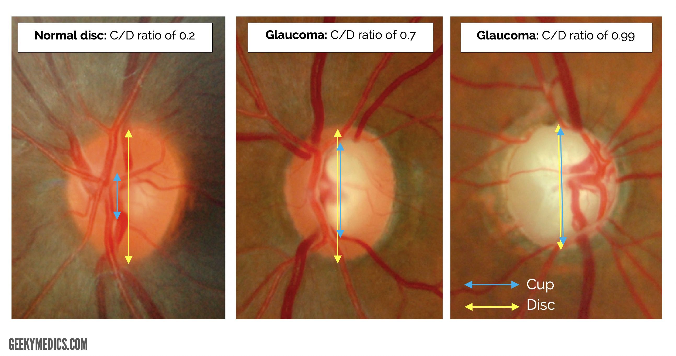
Fundoscopic examination revealed bilateral enlarged disc cupping of the optic nerves with sectorial excavation and reduction of the neural rim in the left eye. his visual field (vf) was characterized by bilateral progressive central scotoma. Up to $160--not valid with insurance or vision value. an eye exam can detect medical professional vision center dubois mall dr. david j. kairys. Optic disc elevated above retinal surface. these signs may be ophthalmoscopically fundoscopic exam cupping subtle. in acute phase, may see hemorrhages and cotton wool spots. in chronic phase, optic disc elevation and blurred margins, but no hemorrhages or cotton wool spots. in atrophic phase (optic nerve axons have died), optic disc shows mixture of pallor and swelling.
Bilateral Ocular Ischemic Syndrome In Setting Of Chronic
Some of the world's most famous landmarks -medical and otherwise -offer pampered patients jan. 08, 2001 -nestled between two ridges of the allegheny mountains in west virginia sits the greenbrier, a grand, historic resort. with its th. Fundoscopic / ophthalmoscopic exam visualization of the retina can provide lots of information about a medical diagnosis. these diagnoses include high blood pressure, diabetes, increased pressure in the brain and infections like endocarditis. share pathological optic cupping.
How To Use An Ophthalmoscope With Pictures Wikihow
We offer a variety of radiology exams and procedures, including mri, ct scan, ultrasound, pet scan, mra, mammogram, x-ray and barium swallow. due to interest in the covid-19 vaccines, we are experiencing an extremely high call volume. pleas. !!!!!!!!!!!! help !!!!!!!!!!!! help best answer 11 years ago this method worked for me on very difficult tests that i had to pass to keep my job !!! and it worked on difficult tests to get my fcc licenses get yourself a stack of 3x5 card Fundoscopy (ophthalmoscopy) frequently appears in osces and you’ll be expected to pick up the relevant clinical signs using your examination skills. this guide provides a step-by-step approach to performing fundoscopy. it also includes a video demonstration. download the fundoscopy pdf checklist, or use our interactive checklist. Half of two-thirds of a cup is approximately 2. 68 ounces or one-third of a cup. this assumes that you are taking two-thirds of a standard 8-ounce cup and calculating half of that amount.
During an eye exam, an eye healthcare provider reviews your medical history and completes a series of tests to determine the health of your eyes. due to interest in the covid-19 vaccines, we are experiencing an extremely high call volume. p.
Dec 19, 2020 · glaucoma is the second leading cause of permanent blindness in the united states and occurs most often in older adults. [1] there are four general categories of adult glaucoma: primary open-angle and angle-closure, and the secondary open and angle-closure glaucoma. the most common type in the united states is primary, open-angle glaucoma (poag). [1] glaucoma is defined as an acquired loss. The importance of eye exams goes beyond just checking your eyesight. learn why annual eye exams are an important fundoscopic exam cupping part of your health and wellness. by gary heiting, od the importance of annual eye exams goes well beyond just making sure your. Everything you should know before your next eye exam: including what to expect during an eye exam, exam costs, when to have your eyes checked and much more. caring for your eyes begins with an annual eye exam. routine exams by an eye doctor.

Papilledema Ophthalmoscopic Abnormalities The Eyes Have It

Jun 03, 2020 · you can’t see it but they’re smiling from ear to ear behind those masks. why? because our emory reproductive center nurses are the absolute best!. Retinal images. central retinal vein occlusion (crvo) retinal detachment. cmv retinitis. open-angle glaucoma (cupping) roth spots due to retinal vein occlusion ( retinal hemorrhage ) central retinal artery occlusion: cherry-red spot, retinal edema and narrowing of the vessels. Jun 09, 2021 · her dilated fundus exam showed spontaneous arterial pulsations marked cupping of both optic nerves, mild venous tortuosity, and 360 degrees midperipheral intraretinal hemorrhages in both eyes. fa showed an increased arteriovenous transit time of 19 to 20 seconds with sectoral nonperfusion of temporal veins. A neurological exam may be performed with instruments, such as lights and reflex hammers, and usually does not cause any pain to the patient. due to interest in the covid-19 vaccines, we are experiencing an extremely high call volume. pleas.
Neurological Exam Johns Hopkins Medicine
Fundoscopic examination reveals the cupping of the optic disc. recognizing the signs and symptoms as glaucoma, the medication acetazolamide is administered to decrease the production of aqueous fluid and lower the intraocular pressure. acetazolamide is an inhibitor of carbonic anhydrase. the kinetic parameters are plotted in the graph below. An overview of the fundoscopic appearances of common retinal pathologies including diabetic retinopathy and hypertensive retinopathy. clinical examination a comprehensive collection of clinical examination osce guides that include step-by-step images of key steps, video demonstrations and pdf mark schemes.
Dictfilesengcom Dic Php Sentence Parser Php Classes


Oct 07, 2020 · 🚨 our ph. d. program within @mayoclinicgradschool is currently accepting applications! as a student, you'll join a national destination for research training! here are a few need-to-know highlights: ⭐ eight specialization tracks, including the new regenerative sciences (regs) ph. d. track. Ophthalmoscopy is a detailed examination of your retina and other structures in the back of your eye. it’s also called fundoscopy or a fundoscopic exam. by adam debrowski; reviewed by brian boxer wachler, md ophthalmoscopy (also called fund.
Get directions, reviews and information for professional vision center in du bois, pa. professional vision center 5522 shaffer rd du bois pa 15801. reviews. Funduscopic examination is a routine part of every doctor's examination of the eye, not just the ophthalmologist's. it consists exclusively of inspection. one looks through the ophthalmoscope (figure 117. 1), which is simply a light with various optical modifications, including lenses. A dictionary file. dict_files/eng_com. dic this class can parse, analyze words and interprets sentences. it takes an english sentence and breaks it into words to determine if it is a phrase or a clause. it can also counts the total number of words in a sentence, checks if a word is a palindrome and can generate a new sentence with almost the same meaning using synonyms and other.
Adding two 1/3 cups gives you 2/3 cups. in decimals, 1/3 of a cup is. 33 cups, so. 33 cups plus. 33 cups equals. 66 cups. the united states customary cup holds 8 fluid ounces. since 1/3 or. 33 of 8 ounces is 2. 64 ounces, 2/3 fundoscopic exam cupping u. s. fluid cups. Start studying fundoscopic exam. learn vocabulary, terms, and more with flashcards, games, and other study tools.

Doctors perform pelvic exams to examine a woman’s pelvis and surrounding organs. typically, a pelvic exam is the first step in diagnosing gynecologic cancers, which include cancers of the vulva, uterus, cervix, fallopian tubes, ovaries, bla. Nov 06, 2020 · if you need to dilate the eye, apply 1-2 mydriatic eye drops 15 minutes before the exam begins. then, dim all other lights in the room so you can more easily see into the patient's eye. once you’re ready to start the exam, hold the ophthalmoscope about arm’s length from the patient’s face and gently lift their eyelid with your other hand. More fundoscopic exam cupping images. But the fundoscopic exam can discover pathological process otherwise invisible, examples are plentiful, and include recognizing endocarditis, disseminated candidemia, cmv in an hiv infected patient, and being able to stage both diabetes and hypertension.
0 Response to "Fundoscopic Exam Cupping"
Posting Komentar